Characterizing the elemental composition of materials is of great interest in scientific disciplines ranging from mineralogy to energy materials. Well established methods like X-ray fluorescence1 provide elemental compositions on the surface and methods such as laser ablation inductively coupled plasma mass spectrometry2 allow isotopic concentration measurements on the surface. Knowledge of the three-dimensional elemental concentration is desirable in many cases and electron- and X-ray based methods were developed for nanometer3 and micrometer4,5 length scales. Destructive methods, analyzing nanometer scale volumes by removing atom by atom, also exist6. All of these techniques require significant sample preparation. While millimeter to centimeter scale 3D visualizations of cracks and average attenuation, without elemental sensitivity, is provided by hard X-ray7,8 or thermal neutron tomography9, a method for three-dimensional element or isotope density measurements on millimeter or centimeter length-scales was hitherto missing.
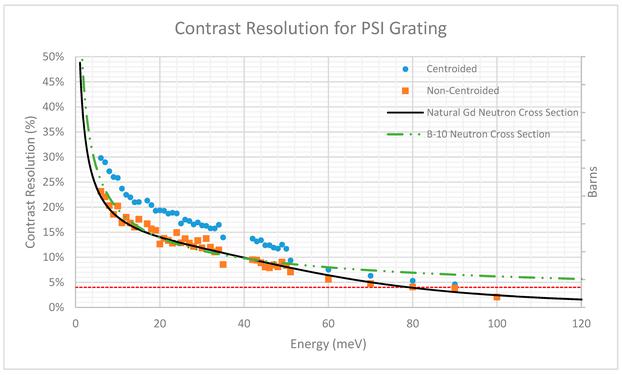
Here, a novel quantitative method is presented based on energy-resolved neutron imaging, utilizing the known neutron absorption cross sections with their ‘finger-print’ absorption resonance signatures. First measurements by Sato et al. showed that neutron absorption resonance spectroscopy with computer tomography can non-destructively provide nuclide density inside an object by analyzing transmitted intensities at absorption resonance energies for specific isotopes10. In this first approach, applicability of the method was limited due to only few projections recorded with low spatial resolution, i.e. 2.3 mm provided by a 64 (8 × 8) pixel 6Li-glass scintillation counter. However, the authors alluded to the potential for quantitative elemental analysis. Utilizing more advanced detector technology, state-of-the-art hardware and software solutions we report, based on 3.25 million state-of-the-art nuclear physics neutron transmission analyses, isotopic densities for five isotopes in 3D in a volume of 0.25 cm3. The method presented in this work is enabled by a pixilated time-of-flight (ToF) neutron transmission detector11 with 512 × 512 pixels and a pixel size of 55 × 55 μm2 installed at an intense short-pulsed spallation neutron source12. Like similar non-isotope specific neutron techniques, the method does not require sample preparation and works with hazardous samples enclosed in containers.
Energy-resolved neutron imaging is often applied to the cold or thermal neutron energy range to gain crystallographic or phase information via Bragg-edge imaging methods13,14,15,16. In this energy range, the attenuation of neutrons by most isotopes is dominated by elastic scattering, limiting the capability for extracting densities reliably, particularly if the material contains multiple elements or the crystals have a preferred orientation (texture). In contrast, utilizing neutron absorption resonances as unique ‘finger-prints’ of isotopes in the epithermal neutron energy range allows to reliably determine the 2D areal-density distribution of isotopes with suitable resonances that can be resolved with the energy resolution provided at the instrument. This is independent of the atomic arrangement, i.e. crystal structure, of the atoms and of the microstructure of the material, i.e. texture, phase fractions etc. Well-developed neutron cross section analysis implemented in state-of-the-art neutron transmission data analysis codes17,18 allows to determine the areal densities of multiple isotopes simultaneously, including considerations such as self-attenuation or resonance interference described by the Reich–Moore formalism19. At a short-pulse neutron source, such as the Los Alamos Neutron Science Center (LANSCE)12, pixilated time-of-flight neutron detectors11 enable therefore three-dimensional isotope density measurements as the number of nuclei of a given isotope can be measured per voxel using tomographic data-sets. Previous research has also estimated the isotope density in two dimensional radiographs20,21, and alluded to the potential of this technique22,23. However, the massive amount of neutron transmission analyses for each pixel recorded in a tomography dataset consisting of several tens or hundreds of rotations, strategies for proper background treatment to obtain reliable results and other obstacles have prevented full development of this intriguing technique. With this work, we demonstrate that three-dimensional isotope density measurements are possible with energy-resolved neutron imaging.
